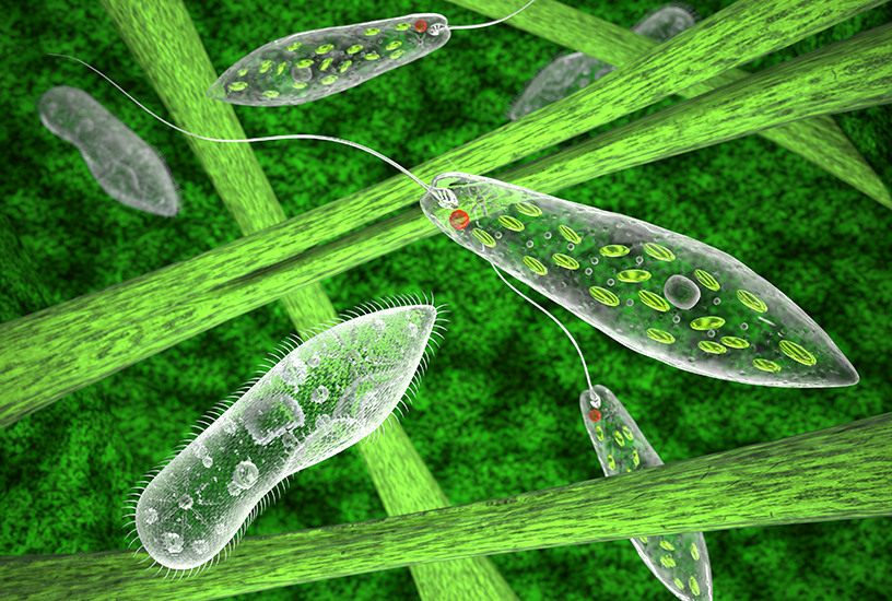
Introduction to Euglena

CURRICULUM ALIGNMENT:
- The theory of evolution by natural selection explains the diversity of living things and is supported by a range of scientific evidence
- Describing biodiversity as a function of evolution
BACKGROUND:
Euglena are classified under the Kingdom Excavata and many species possess chloroplasts. However, unlike plant chloroplasts, which are enclosed by two membranes, the Euglena chloroplasts are surrounded by three membranes. The presence of three membranes, along with molecular evidence, indicates that Euglena chloroplasts emerged from a secondary endosymbiotic event, whereby a unicellular green alga was engulfed by a Euglenoid. Euglena move by whipping their flagellum in a propelling motion. Protoslo makes this movement easier to observe as it slows down the Euglena by increasing the viscosity of the water. It also changes the refractive index of water just enough to provide increased contrast with the flagellum, making it more visible.
This practical provides an excellent opportunity for students to observe a microscopic organism, understand its physiology, identify the unique traits it exhibits and discover how their form relates to their function. Students are tasked with observing Euglena under the microscope and identifying the chloroplasts, pyrenoids, contractile vacuole, eyespot (stigma), nucleus and flagellum. Students will also apply Protoslo to the microscope slide to observe the movement of the flagellum more easily. This practical provides a great introduction into microscopic organisms, basic lab observation practices, and allows students to gain a deeper understanding of their evolutionary trait
PREPARATION - BY LAB TECHNICIAN
Preparing the Culture- Loosen the lid of the Euglena culture as soon as it arrives and place it on a flat surface with access to natural light.
- When ready to use the culture, remove the lid and aerate the culture using a transfer pipette.
- Allow the culture to rest for 5 to 15 minutes, and then examine it with a stereomicroscope at 20 to 40X.
- Identify areas of Euglena concentration and instruct students to remove their samples from these areas.
Preparing Workstations
- Provide each workstation with the following materials.
- Euglena Culture
- Protoslo Solution
- Transfer Pipette
- Microscope Slides
- Coverslips
- Microscope
METHOD - STUDENT ACTIVITY
- Collect the Euglena culture and bring it to your workstation.
- Observe the culture with the naked eye and note what you see.
- Remove a sample from the culture jar, using a transfer pipette by compressing the bulb of the pipette between your thumb and forefinger and lowering the tip to the bottom of the culture jar. Then, gently release the pressure on the bulb and allow the sample to be sucked into the pipette. It is best to remove the sample from the bottom of the jar, as this is where the Euglena will be in the greatest concentration.
- Deliver 2–3 drops of your sample onto a microscope slide.
- Add a drop of Protoslo along with your drops of culture and thoroughly mix on a microscope slide.
- Carefully place a coverslip over the top.
- Observe the sample under your microscope on a low-power objective. Search the water for swimming spindle-shaped cells.
OBSERVATION AND RESULTS
Students should observe:- Chloroplasts: Green structures that contain the pigment Chlorophyll. These should be seen in abundance. The prevalence of these chloroplasts can even make observation of other organelles difficult.
- Pyrenoids: These food storage bodies can be identified as dots near the centre of each chloroplast.
- Contractile vacuole: Frequently expanding to a larger size and collapsing suddenly, the contractile vacuole is a clear spherical structure that contracts by discharging its contents into the surrounding medium.
- Eyespot (stigma): The eyespot is found near the anterior end and appears red in colour.
- Nucleus: The Euglena nucleus is located roughly in the centre of the cell and contains a darker body; known as the endosome. It is best to view these two elements using stained preparations.
- Flagellum: Used for locomotion, the flagellum is a whip-like organelle located at the anterior end. Students should observe the movement of the flagellum.
INVESTIGATIONS
- Challenge students with identifying one characteristic Euglena have in common with animals, and one they have in common with plants.
- Provide students with a piece of paper to draw a Euglena and label the parts.
TEACHER TIP:
You may wish to use a prepared Euglena microscope slide to allow students to observe the nucleus and endosome in more detail
 Time Requirements
- 45 mins
![]() Material List
Material List
- Euglena Culture
- Protolo Solution
- Plain Microscope Slides
- Transfer Pipettes
- Coverslips
- Petri Dishes
- Stereo Microscope
- Compound Microscope
 Complimentary products
 Safety Requirements
- Wear appropriate personal protective equipment (PPE).
-
Wash your hands thoroughly before and after the practical.
- Do not release any organisms into the environment.
- Disinfect work areas before and after the practical.
